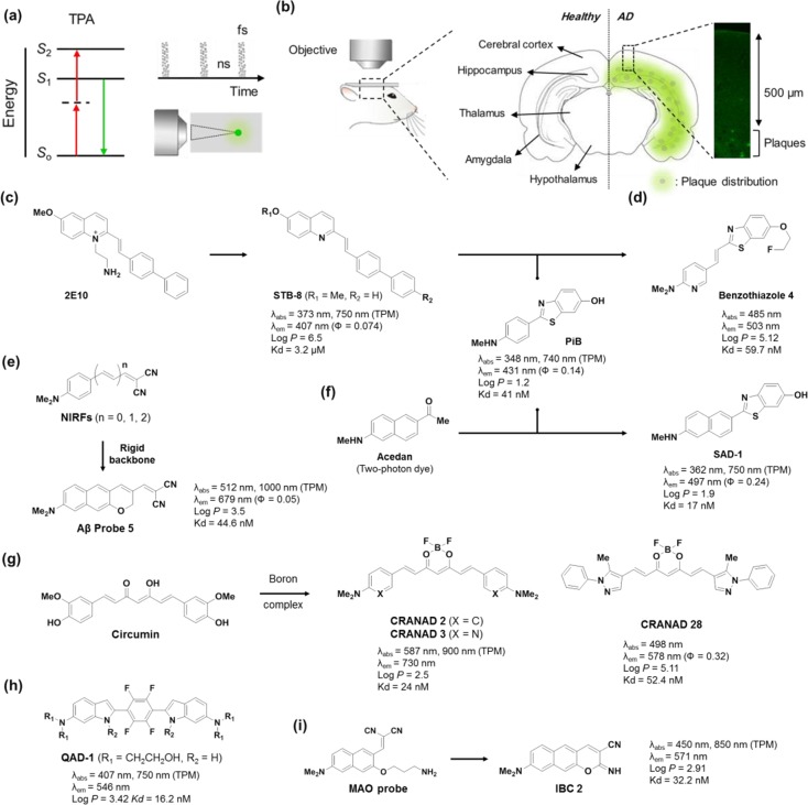
Figure 3.
Two-photon probes for detecting AD biomarkers. (a) Illustration of two-photon absorption, pulsed laser and focal point excitation. (b) Illustration of the distribution of amyloid-β plaques in the brain. (c–h) Two-photon amyloid-β probes: (c) 2E10, STB-8, PiB, (d) benzothizole 4, (e) NIRFs, Aβ probe 5, (f) SAD-1, (g) CRANADs, and (h) QAD-1. (i) A two-photon dual probe for MAOs and amyloid-β plaques.

