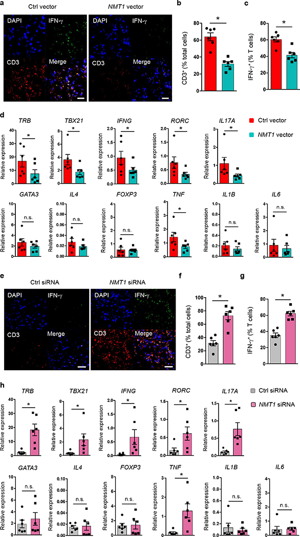
Fig. 3. N-myristoylation protects against synovial inflammation.

(a-d) RA CD4+CD45RO– T cells were transfected with an NMT1 or control expression vector and injected into NSG mice engrafted with human synovial tissue. Synovial tissues were harvested for transcriptome analysis and immunostaining. (a) Tissue sections stained with anti-IFN-γ (green) and anti-CD3 (red). Nuclei marked with DAPI. Representative images from 6 grafts. Scale bars 20 μm. (b, c) Frequencies of CD3+ T cells and of CD3+IFN-γ+ T cells in tissue sections. (d) Gene transcripts measured in tissue extracts by qPCR. (e-h) Healthy CD4+CD45RO– T cells were transfected with NMT1 siRNA or control siRNA and adoptively transferred into NSG mice engrafted with human synovial tissue. Synovial explants were analyzed for gene expression and by immunohistochemical staining. (e) Tissue-infiltrating human T cells evaluated by dual-color immunostaining of CD3 (red) and IFN-γ (green). Nuclei marked with DAPI. Representative images from 6 grafts. Scale bars 20 μm. (f, g) Frequencies of tissue-residing CD3+ T cells and CD3+IFN-γ+ T cells. (h) Tissue transcriptome of inflammation-associated genes determined by qPCR.Data are mean ± SEM from 6 synovial grafts. Paired Mann-Whitney-Wilcoxon rank test. *p < 0.05.
