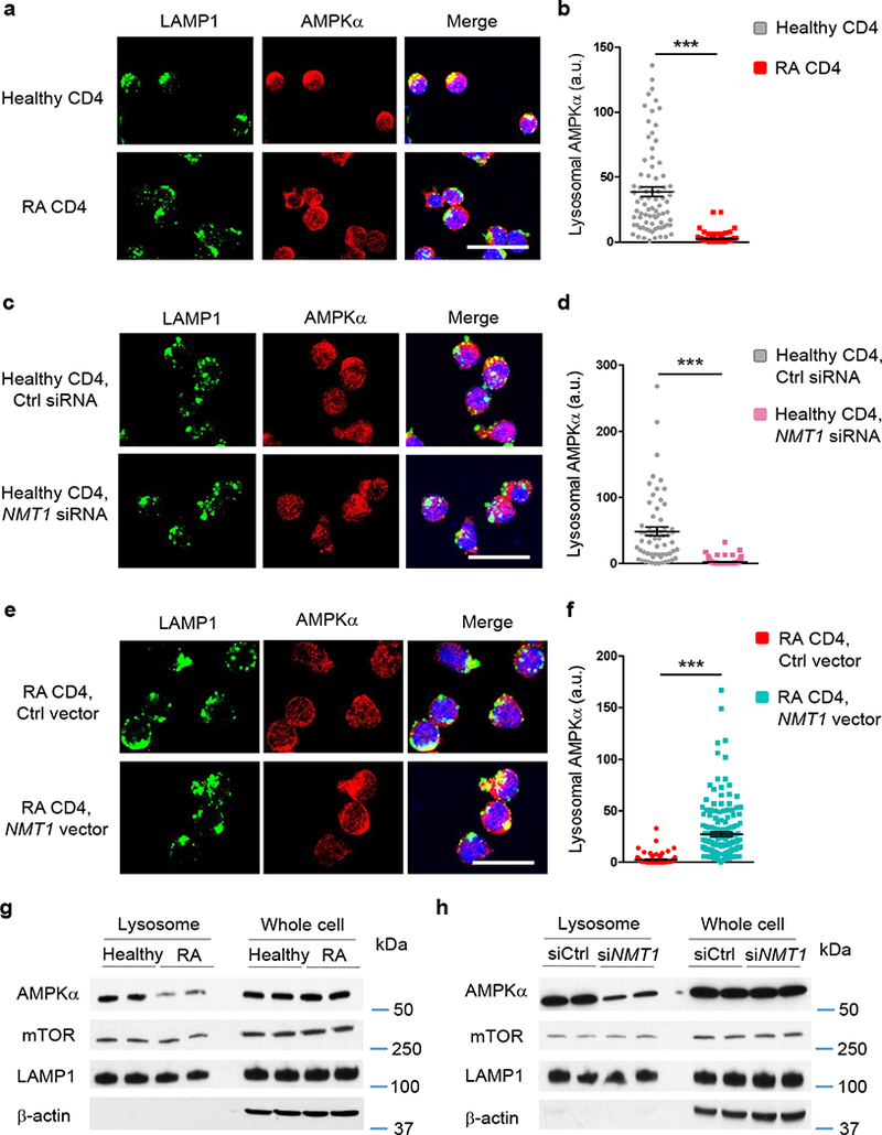
Fig. 6. NMT1 controls lysosomal recruitment of AMPK.

CD4+CD45RA+ T cells from RA patients and healthy individuals were stimulated for 72 h. (a-f) Lysosome was identified by staining with rabbit anti-human LAMP1 (green). AMPKα was localized with mouse anti-human AMPKα (red). Individual cells were analyzed for LAMP1-AMPKa co-localization by confocal microscopy and each dot represents one T cell. Scale bars 20 μm. (a, b) Lysosomal localization of AMPK in RA and control T cells. Representative immuno-stains and quantification of the co-localization signal in individual cells. Data are from 6 RA patients and 6 healthy controls. (c, d) T cells from 6 healthy individuals were transfected with NMT1 siRNA or control siRNA and stimulated for 72 h. (e, f) T cells from 6 RA patients were transfected with a NMT1 expression vector or a control vector and stimulated for 72 h. Data (mean ± SEM) collected from 6 samples in each group. ***p < 0.001 by unpaired Mann-Whitney-Wilcoxon rank test. (g) Lysosomes were isolated from stimulated CD4+ T cells and immunoblotted for AMPKα and mTOR. Whole cell lysates were used as controls. Representative immunoblots from a total of 4 RA patients and 4 healthy controls. (h) CD4+CD45RA+ T cells from healthy individuals were transfected with NMT1 siRNA or control siRNA and stimulated for 72 h. AMPKα and mTOR expression was quantified in lysosomal fractions and in whole cell lysates. Shown are representative immunoblots from a total of 4 individuals.
