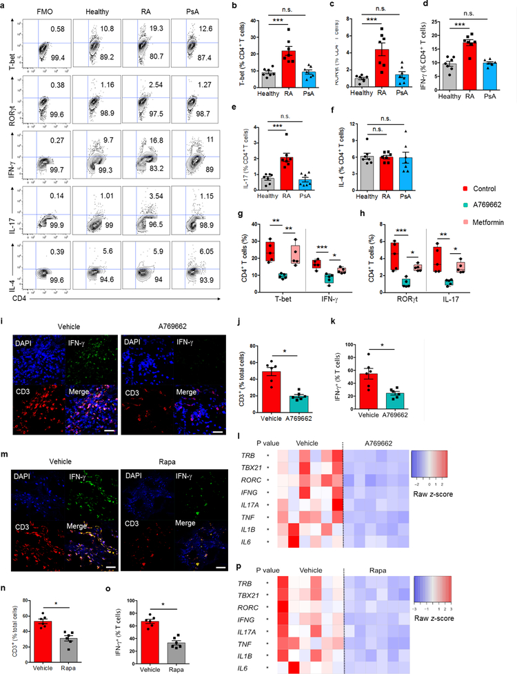
Fig. 8. AMPK activation corrects arthrogenic T cell effector functions in vitro and in vivo.
CD4+CD45RA+ T cells from RA patients, PsA patients and healthy controls were stimulated for 4 days and expression of the lineage-determining transcription factors T-bet and RORγt was assessed by flow cytometry. On day 6 after stimulation, intracellular cytokines (IFN-γ, IL-17, IL-4) were quantified. All data are mean ± SEM. (a-f) Representative contour plots and cumulative data from 7 patient-control pairs. One-way ANOVA and post-ANOVA pair-wise two-group comparisons with Tukey’s method. (g, h) RA T cells were stimulated in the absence or presence of the AMPK activators A769662 (10 μM) or metformin (50 μM). T-bet and RORγt were analyzed on day 4, IFN-γ and IL-17 on day 6. Median frequencies and interquartile ranges are indicated. One-way ANOVA and post-ANOVA pair-wise two-group comparisons were conducted with Tukey’s method. (i-p) NSG mice were engrafted with human synovial tissue and reconstituted with PBMCs from RA patients. Such chimeras were treated with vehicle (i-p), A769662 (i-l) or rapamycin (m-p). Tissue transcriptomes and tissue T cells were analyzed in synovial explants after 7 days of treatment. (i, m) T cell infiltrate in the synovial tissue shown by dual-color immunostaining of CD3 (red) and IFN-γ (green). Nuclei marked with DAPI. Representative images from 6 grafts. Scale bar 20 μm. (j, k and n, o) Frequencies of tissue CD3+ T cells and of CD3+IFN-γ+ T cells. (l, p) Tissue transcriptome of inflammation-associated genes from 6 synovial grafts in each group. Paired Mann-Whitney-Wilcoxon rank test. *p < 0.05. **p < 0.01. ***p < 0.001.

