Figure 2.
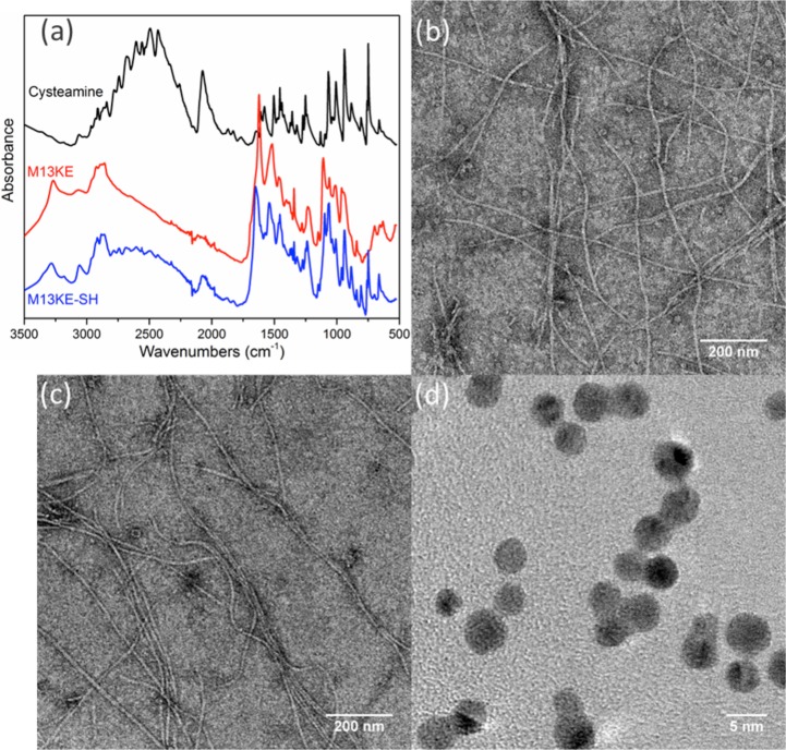
Preparation of thiolated phage capsids and AuNPs. (a) ATR-FTIR of purified thiolated M13KE phage indicates gain of S–H stretching (2550 cm–1) and C–S stretching (659 cm–1) signals. Shown are cysteamine (black), phage before modification (red), and phage after modification (blue). Representative TEM images of wild-type M13KE phage (b) before and (c) after thiolation indicate little change in gross morphology. (d) TEM image of AuNPs shows homogeneous particles of ∼4 nm diameter.

