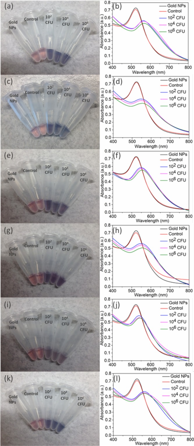
Figure 5.

Detection of several bacterial species in relevant media: (a, b) V. cholerae 0395 in seawater, (c, d) X. campestris (pv campestris) in tap water, (e, f) X. campestris (pv vesicatoria) in tap water, (g, h) P. aeruginosa in tap water, and (i, j) human serum and (k, l) E. coli (I+) in tap water. The corresponding chimeric phage (Table 1) was used in each case. Shown are digital photographs (left) and UV–vis spectra (right). Left column: from left to right, samples contain AuNPs and no bacteria or phages (black line in right column), unmodified phage with 106 CFU host bacteria (red line in right column), and thiolated phage with host bacteria at 102, 104, and 106 CFU (blue, magenta, and green lines, respectively, in the right column).
