
Figure 2.
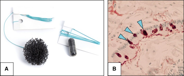
Cytosponge oesophageal cell collection device. (A) Cytosponge oesophageal cell collection device in a gelatin capsule (right) and expanded (left). (B) Trefoil‐factor 3 staining (20×) from a patient with Barrett's oesophagus showing columnar lined epithelium with goblet cells (arrowheads). Courtesy of Dr Maria O'Donovan (Department of Histopathology, Cambridge University Hospitals NHS Foundation Trust, Cambridge, UK).