Figure 5.

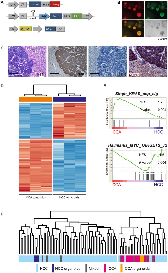
Liver organoids retain their plasticity to differentiate into tumors resembling HCC. (A) Liver organoids derived from WT C57Bl/6J mice were stably transduced with retroviruses expressing Myc coupled with a red fluorescent protein (mCherry), a GFP‐coupled shRNA directed against p53, and transiently transfected with pX459_sgAPC. (B) Stable integration of both retroviral vectors is indicated by expression of fluorescent markers, GFP and mCherry. (C) Myc‐driven liver tumors develop at a latency of about 40 days and are histologically distinct from Kras‐driven murine CCAs. Tumor cells stain negative for CK19 and strongly express glutamine synthetase. Immunohistochemistry confirms GFP positivity. Scale bars represent 200 µm unless otherwise indicated. (D) Unsupervised cluster analyses based on differentially expressed genes between HCC and CCA tumoroids based on Euclidean distance. (E) Gene‐set enrichment analyses of key gene sets activated in CCA and HCC tumoroids. (F) Integrative analyses of a publicly available cohort of 70 human HCCs, 17 CCAs, and 7 liver cancers of mixed HCC/CCA histology, with Myc HCC‐like and KrasG12D/wt CCA tumoroids. Integration and cross‐species comparisons were performed as described by Lee et al.26 Unsupervised cluster analyses confirm that the HCC and CCA tumoroids share gene‐expression profiles with authentic human cancers. Abbreviations: Mixed, liver cancers of mixed HCC/CCA histology; and NES, normalized enrichment score.
