Fig. 3.
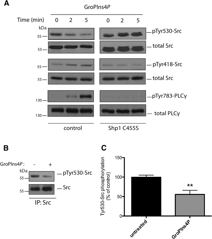
GroPIns4P induces Src dephosphorylation. a Representative western blots using anti-phosphotyrosine 530 in Src (pTyr530-Src), anti-phosphotyrosine 418 in Src (pTyr418-Src) and anti-phosphotyrosine 783 in PLCγ (pTyr783-PLCγ) specific antibodies in serum-starved NIH3T3 cells non-transfected or over-expressing the dominant negative Shp1-C455S mutant and treated with 50 μM GroPIns4P for the indicated times (see the top of the two panels). Total Src and total PLC were used as loading controls. Data are representative of three independent experiments. Molecular weight standards (kDa) are indicated on the left of each panel. b Representative immunoprecipitated Src fraction (IP: Src) from NIH3T3 cell lysates washed and incubated with purified recombinant Shp1 for 10 min at 37 °C in the absence (−) or presence (+) of 50 μM GroPIns4P (as indicated). The top panel shows western blots with an anti-phosphotyrosine antibody (pTyr530-Src) to reveal the specific phosphorylation of Tyr-530 in Src. The blot was then re-probed with an anti-Src antibody for immunoprecipitated proteins (bottom panel). Molecular weight standards (kDa) are indicated on the left of each panel. c Quantification of Src phosphorylation in samples treated with GroPIns4P (as in b) by the ImageJ analysis software. Data (GroPIns4P) are expressed as percentages of untreated sample (untreated) of the means (±SD) of three independent experiments, each of which was performed in duplicate (n = 6). **P < 0.02 (Student’s t-test)

