Fig. 5.
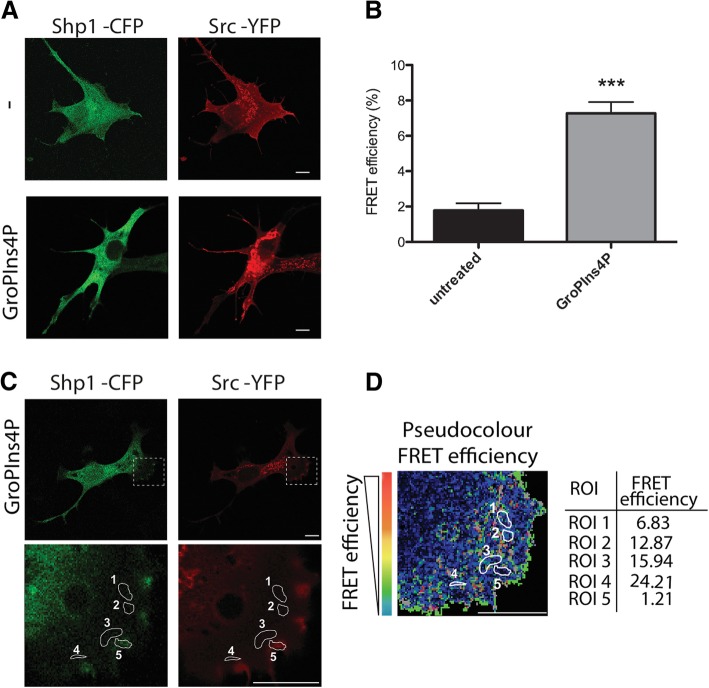
GroPIns4P promotes the association of Shp1 and Src within the lamellar region. a Representative confocal microscopy images of serum-starved NIH3T3 cells co-transfected with Shp1-CFP (green) and Src-YFP (red) for 24 h and then untreated (−) or treated with 50 μM GroPIns4P (GroPIns4P) for 5 min and subjected to Acceptor Photobleaching apFRET analysis. b Quantification of the FRET efficiency over the cells treated as in a. c Representative confocal microscopy images of serum-starved NIH3T3 cells co-transfected with Shp1-CFP (green) and Src-YFP (red) for 24 h and then treated with 50 μM GroPIns4P for 5 min before apFRET analysis. The dashed rectangle in the lamellar region (close to the plasma membrane) indicates where the FRET efficiency was analysed. d Representative colour-coded apFRET efficiency of the lamellar region (selected in a) where the colour-scale quantifies the degree of protein-protein interaction in each of the indicated regions of interest (ROIs 1 to 5, white circles; the FRET efficiency for each ROI is reported in the table on the right). Data are expressed as the means (±SD) (n = 15 cells/condition). ***P < 0.005 (Student’s t-test). Scale bars, 10 μm

