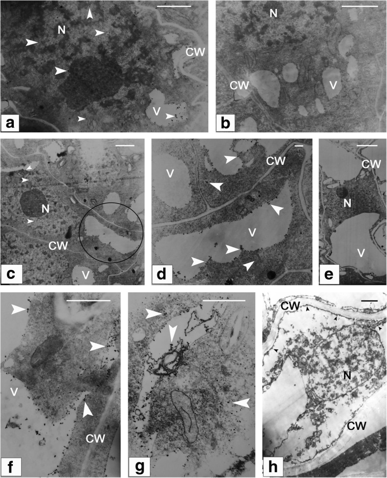
Fig. 9.

Subcellular localization of calcium within the nucellar cells involved in pollen chamber formation. (a) At the early stage of ovule development, a relatively higher Ca2+ precipitation is found in both of vacuoles and nucleus, compared with that in the cytoplasm. (b) A negative control of (a) without Ca2+ particles. (c), (d), (f), and (g) The nucellar cells at the micropylar end become elongated in shape, and distribution of Ca2+ precipitation is found to be increased in these nucellar cells. (d) Magnified view of the circled area in (c) shows numerous Ca2+ particles within the vacuole and cytoplasm. (e) A negative control of (c) without Ca2+ particles. (g) Deformed endoplasmic reticula are enclosed within the vacuole. (h) Few of Ca2+ particles are distributed along the cytoplasm debris, with no detectable aggregation of Ca2+ in the nucleus of the dying nucellar cell. Arrows indicate Ca2+ particles. Bars in (a), (b), (c), (e), and (h) = 2 μm, and in (d), (f), and (g) = 0.2 μm. CW, cell wall; N, nucleus; V, vacuole
