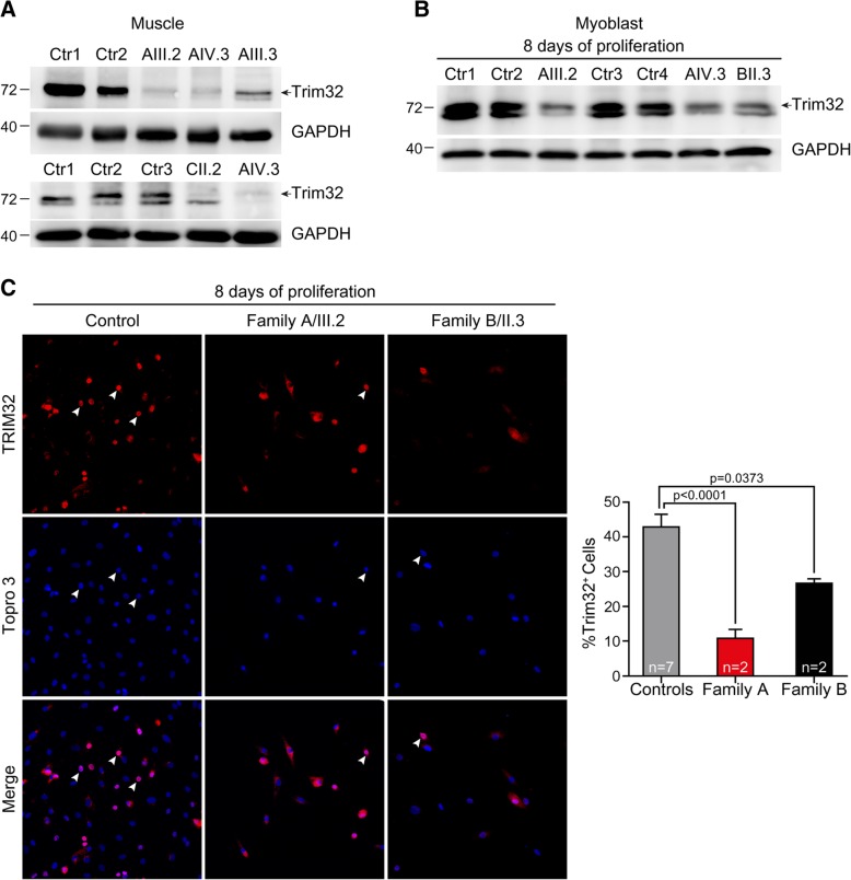
Fig. 2.
Reduced TRIM32 protein level associated with p.V591 M, p.N217S/F568del and p.C39LfsX17 mutations. a, b Western blot analysis of a muscle derived from biopsies of family A patients, family C patient and healthy controls, and b primary myoblasts from family A patients, family B patient, and healthy controls. Anti-TRIM32 antibody revealed a remarkable reduction of TRIM32 protein level in TRIM32V591M, TRIM32N217S/F568del and TRIM32C39LfsX17 samples compared with controls. An anti-GAPDH blot is shown as a loading control. Arrows indicate the correct band at 72 kDa. c. Immunofluorescence staining and quantification of the percentage of TRIM32+ cells (arrowheads) in primary myoblasts at 8 days growing in proliferation medium from family A patients (n = 2), family B patients (n = 2) and healthy controls (n = 7). Images show TRIM32 fluorescence (red). Cells were counterstained with Topro 3 (blue) to visualize the nuclei. The percentage of TRIM32+ cells was significantly lower in TRIM32V591M and TRIM32N217S/F568del myoblasts than in controls. Data from 9 to 31 independent fields were analyzed per condition. Mean ± SEM; Kruskal-Wallis with Dunn’s multiple comparison test. Scale bar, 50 μm

