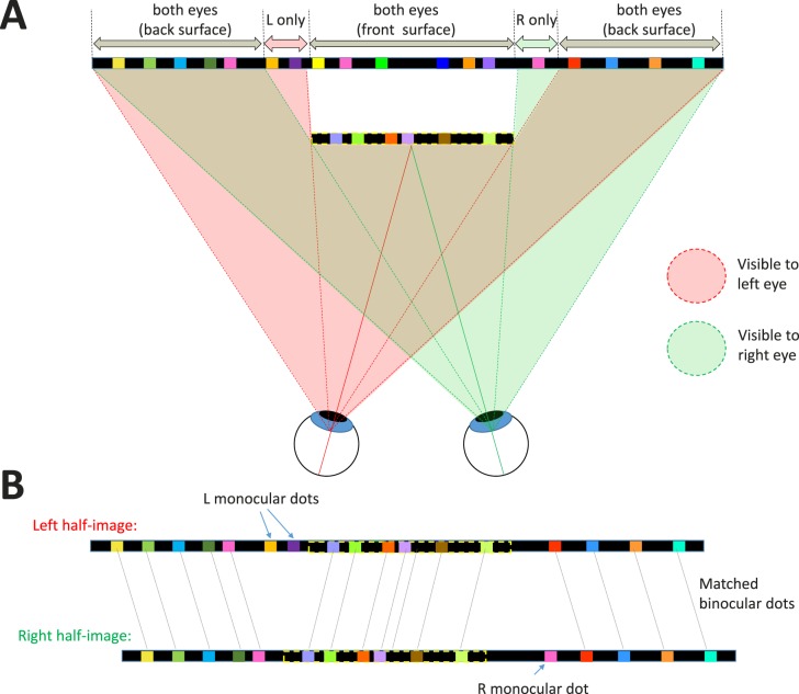
Figure 4.
Binocular occlusion geometry for our stimulus where a target surface (outlined in dashed yellow lines) appears in front of a background. (A) The front surface occludes different regions of the background in the two eyes. The pink (green) shaded regions indicate which parts of the stimulus are visible to the left (right) eye, respectively. To the left of the front surface, there is a narrow strip of the back surface that is visible only to the left eye. Dots in this region are accordingly left-monocular, visible only to the left eye. The same applies for the right eye, for a strip to the right of the front surface. (B) Shows the resulting left- and right-eye half-images. Most dots are binocular, that is, visible in both eyes, so a disparity can be defined. Dots on the back surface have uncrossed disparity (position in the left half-image is to the left of position in the right half-image), whereas dots on the front surface have crossed disparity (position in the left half-image is to the right of position in the right half-image). The monocular dots have no matching dots in the other eye and are said to be uncorrelated. Nothing identifies the uncorrelated dots in either half-image individually; they can be detected only when the two eyes' half-images are compared.

