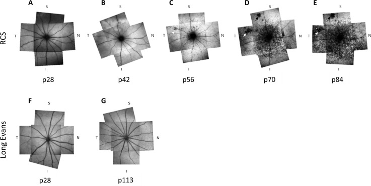
Figure 1.
BL-FAF longitudinal follow-up in a representative RCS rat. BL-FAF imaging was performed at indicated days postnatal in a representative RCS rat (A–E). Each panel is a composite of five BL-FAF scans. To generate the composite image, the size of the optic disc was matched and then the images were aligned according to the blood vessel orientation. An ImageJ plugin (“Align images by line Region of Interest [ROI]”) was used to align the composite images. (F, G) BL-FAF scans of two representative Long Evans rats at ages p28 (F) and p113 (G). T, temporal; N, nasal; S, superior; I, inferior.

