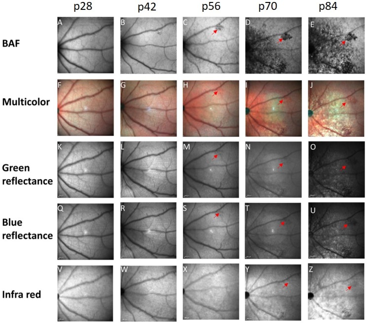
Figure 6.

Comparison between different fundus imaging modalities. A representative example of a single-animal follow-up at ages p28 to p84 using BL-FAF (A–E), multicolor fundus imaging (F–J), green reflectance (K–O), blue reflectance (Q–U), and IR fundus imaging (V–Z). A red arrow highlights the same hypofluorescent lesion in the different scans.
