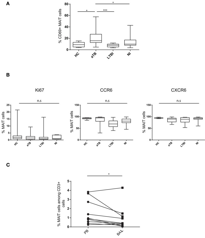
Figure 2.
MAIT cells are activated in the peripheral blood and do not accumulate in the lung in children with active TB. (A) box and whisker plots show median, interquartile range and the minimal to maximal proportions of MAIT cells expressing CD69 (Mann-Whitney test). (B) box and whisker plots show median, interquartile range and the minimal to maximal proportions of MAIT cells expressing the indicated marker in the different children groups. Differences were not significant (Mann-Whitney test). (C) correlation of MAIT cell frequencies in the peripheral blood (PB) and bronchoalveolar lavage (BAL) in aTB children (Wilcoxon matched-pairs signed rank test). Each point corresponds to 1 patient and lines connect matched samples. *P < 0.05; ***P < 0.0001, n.s, not significant.

