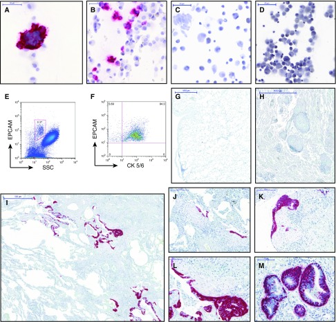
Figure 4.
Increase in CK5/6+ ∆NP63+ airway basal cells (ABCs) in BAL and alveolar tissue of patients with idiopathic pulmonary fibrosis (IPF). (A–D) CK5/6+ ABC (stained in red) were found frequently in cell smears of BAL from patients with IPF (A and B) and often formed cell clusters. In contrast, CK5/6+ ABC were found only rarely in the BAL of old healthy volunteers (C) and patients with chronic obstructive pulmonary disease (D). Flow cytometry revealed presence of EPCAM+ epithelial cells in the BAL of patients with IPF, and most of them coexpressed CK5/6, identifying these cells as ABC (E and F). Immunohistochemistry of lung tissues stained for CK5/6 (red) and ∆Np63 (nuclear turquois) showed ABCs in the basal layer of airway epithelium but not in the alveolar compartment of normal lung tissue (G) and sarcoid tissue (H). In contrast, in IPF tissues we observed an enrichment of ABCs within the alveolar compartment (I–M). ABCs frequently covered fibroblast foci (J–L), and occasionally formed hollow structures (M). In some patients, alveolar epithelium was replaced by multiple layers of ABCs consistent with basal cell hyperplasia and squamous metaplasia (L and M). Scale bars: A–D, 50 μm; G and H, 1,000 μm; I, 500 μm; J and K, 200 μm; L and M, 100 μm. CK5/6 = cytokeratin 5/6; SSC = side scatter.

