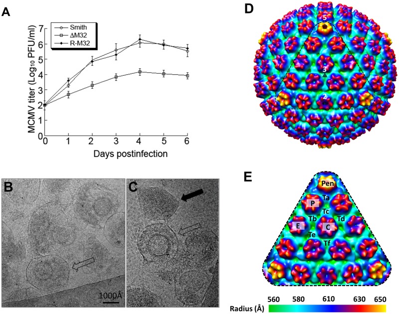
Fig 9. Generation and analyses of M32-deletion MCMV mutant.
(A) Growth of the parental virus MCMVBAC (Smith), M32-deletion mutant ΔM32 and rescued mutant R-M32 in NIH 3T3 cells. NIH 3T3 cells were infected with each virus at a MOI of 0.5 PFU per cell. At 0, 1, 2, 3, 4, 5, and 6 days post-infection, we harvested the cells and culture media and determined the viral titers by plaque assays on NIH 3T3 cells. The error bars indicate the standard deviations based on triplicate experiments. (B-C) CryoEM images of the ΔM32 MCMV mutant. Fully enveloped particle and dense body are indicated by an open and a solid black arrow, respectively. (D) Radially colored surface representation of cryoEM reconstruction (~25 Å resolution) of the enveloped particles of ΔM32 MCMV, viewed along a 3-fold axis. The pentagon, triangle, and oval symbol denotes a 5-fold, 3-fold, and 2-fold axis, respectively. (E) Enlargement of a facet [triangle region in (D)] with pentons (Pen), hexons (C, E, and P) and triplexes (Ta, Tb, Tc, Td, Te, and Tf) indicated.

