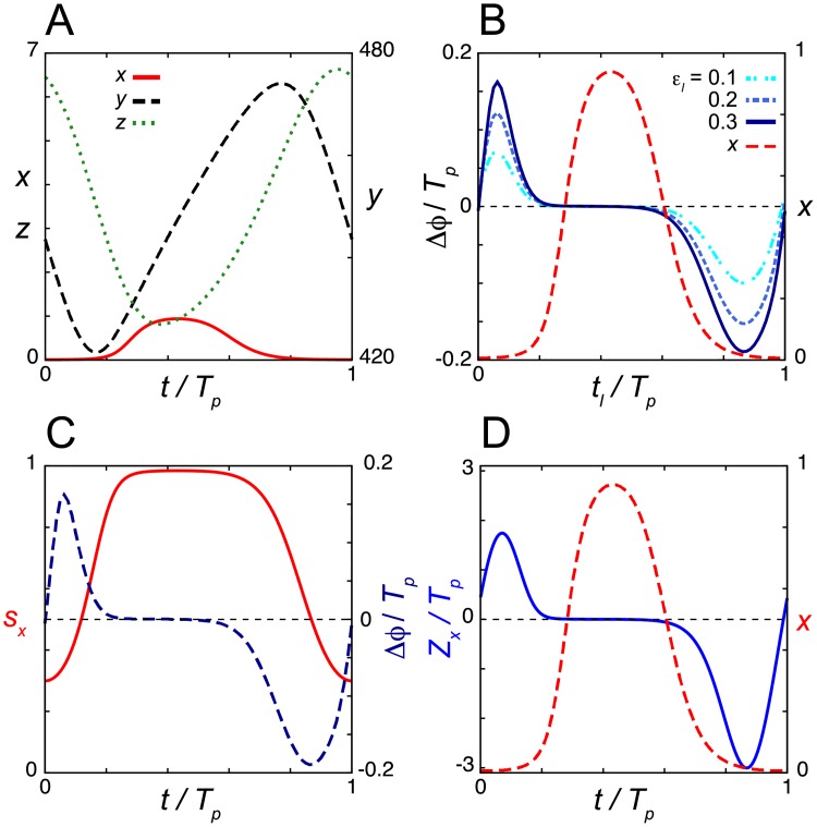
Fig 5. Dead zone generated by the saturation of repressor translation in the induction response.
(A) Time series of the levels of mRNA x, cytoplasmic protein y and nuclear protein z in Eqs (8), (10) and (11) in the absence of a light signal. (B) Phase shift Δϕ as a function of the onset of light signals tl. Results for different values of εl are shown. (C) Time series of the saturation index sx = x/(Kt+x). The PRC with εl = 0.3 is also plotted (right y-axis). (D) Phase sensitivity Zx. In (B) and (D), the time series of x (red broken line) is plotted (right y-axis) as a reference. Values of reaction parameters are listed in S1 Table. Tp = 24. In (B), Td = 0.5Tp/24 = 0.5.

