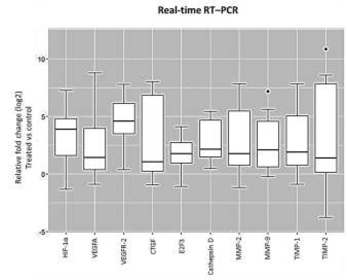
Figure 5.
Box plot analysis of hypoxia-inducible factor 1 (HIF-1α), vascular endothelial growth factors A (VEGFA), vascular endothelial growth factors receptor 2 (VEGFR-2), and connective tissue growth factor (CTGF), E2F3, cathepsin D, matrix metalloproteinase-2 (MMP-2), MMP-9, tissue inhibitors of metalloproteinases 1 (TIMP-1) and TIMP-2 expression in retinal pigment epithelial (RPE) cell cultures exposed to extremely low frequency-pulsed electromagnetic fields (ELF-PEMF). Cultures exposed to ELF-PEMF and the cells without exposure were considered as treatment and the control, respectively. After 3 days, RNA was extracted, and gene expression analysis was performed with quantitative real-time RT–PCR as described in the methods section. mRNA levels were normalized to Glyceraldehyde 3-phosphate dehydrogenase (GAPDH) and presented as log2 fold change of the control values. ELF-PEMF increased gene expression of HIF-1α, VEGFA, VEGFR-2, CTGF, E2F3, MMP-2, MMP-9 and TIMP-1 in treated cell cultures compared to the control (P<0.05). However, gene expression of cathepsin D and TIMP-2 were not altered (P>0.05)

