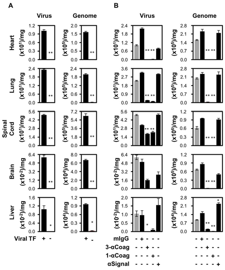
Fig. 1.

Infection of Th2 immuno-dominant mice (BALB/c) is enhanced by TF on HSV1. (A) Eight-week-old female BALB/c mice were inoculated intravenously with 5 × 10⁵ PFU of either TF competent (TF+; n=24) or deficient (TF−; n=13) HSV1 via the tail vein. Three days post-infection, the mice were processed and the amount of infectious virus (Virus; PFU/mg) and HSV1 genetic material (Genome; genome copies/mg) were determined. (B) Additional experiments were conducted after pre-immunization of mice with a mixture of three anti-TF IgG1 monoclonal antibodies, 5G9, 9C3 and 6B4 (3-αCoag; at 0.33 mg each/mouse; n=10), 5G9 alone (1-αCoag; 1 mg/mouse; n=6) or 10H10 alone (αSignal; 1 mg/mouse; n=6), 4 hours prior to injection of the virus. Purified anti-Simian virus 40 large T antigen (mIgG; 1 mg/mouse; n=8) was used as an irrelevant monoclonal IgG1 subclass-matched negative control. Grey bars represent data from HSV1/TF+ as depicted in Panel A. In all panels data are expressed as mean ± SEM. As determined by Student’s t test, **P ≤ 0.05 and *P ≤ 0.10 when compared to the TF+ virus.
