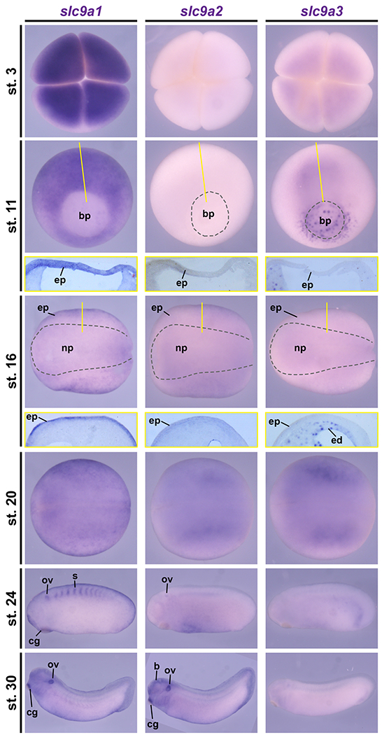
Figure 1: Dynamic slc9a1, 2 and 3 expression during Xenopus development.

In situ hybridization on Xenopus laevis embryos from stages (st.) 3 – 30 revealed dynamic expression of slc9a transcripts during early development. At the four-cell stage (st. 3) strong maternal transcript deposition was detected for slc9a1, but slc9a3 was only lowly expressed and no signals were observed for slc9a2. During gastrulation (st. 11), slc9a1 was markedly expressed throughout the prospective ectoderm and mesoderm, while slc9a3 was found at the blastpore (bp), the endoderm as well as involuting mesodermal cells and no specific expression was found for slc9a2. In neurula stage embryos (st. 16 and 20), slc9a1 and 2 transcripts were detected in the epidermal ectoderm (ep) with stronger expression of slc9a1, and slc9a3 was expressed in the endoderm (ed). During late neurulation (st. 20), slc9a2 and 3 were also expressed in the presomitic mesoderm. In tailbud stages (st. 24, 30), slc9a1 and 2 transcripts were detected in the otic vesicle (ov) and the cement gland (eg), but not slc9a3. Additionally, slc9a1 was expressed in the somites (s), and slc9a2 was expressed in the brain (b). St. 3 embryos are depicted in animal view. St. 11 embryos are depicted in vegetal view, dorsal up. St. 16 and 20 embryos are depicted in dorsal view, anterior left. St. 24 and 30 embryos are depicted in lateral view, anterior left. Sections depicting the epidermis of st. 11 and st. 16 embryos are shown in yellow boxes below the whole mount image. Section planes are indicated by yellow lines.
