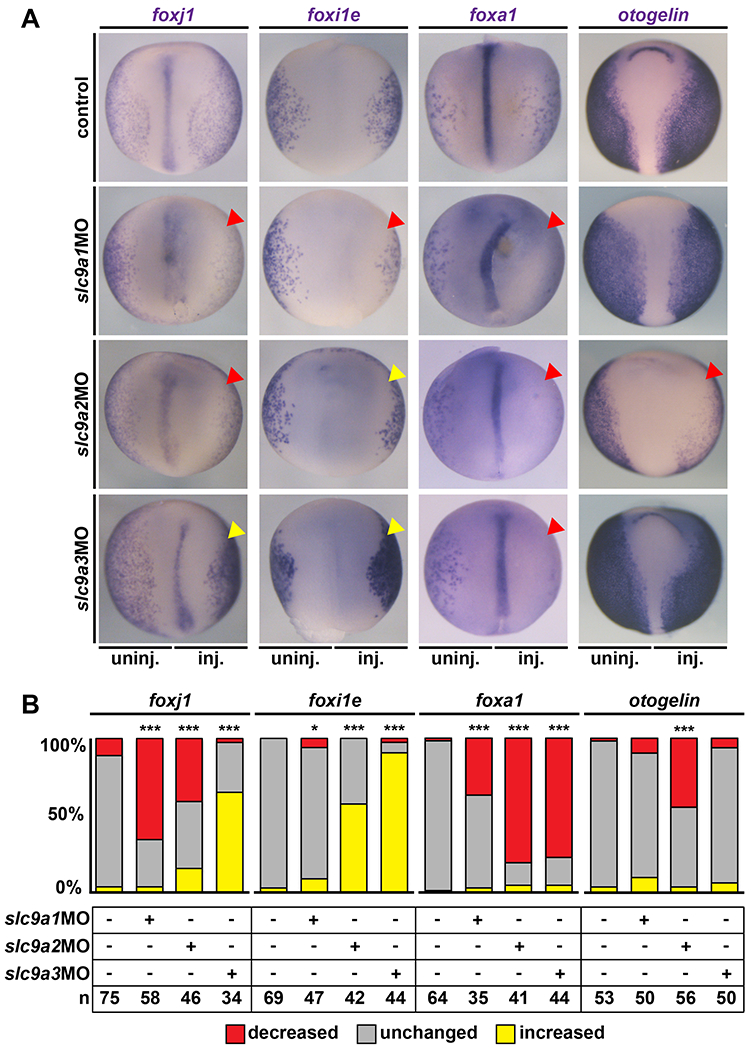
Figure 4: NHE1-3 Ioss-of-function affects gene expression in MCCs and secretory cells of the mucociliary epidermis.

In situ hybridization for cell type markers in the embryonic mucociliary epidermis. Embryos were injected unilaterally on the right side (inj.) and the uninjected left side (uninj.) served as internal control. Unmanipulated specimens were used as additional controls (control). (A) St. 17 embryos are shown in dorsal view, anterior up. foxj1 marks MCCs, foxi1e marks ion secreting cells, foxa1 marks small secretory cells and otogelin is a marker of mucus secreting goblet cells. Downregulation is indicated by red arrowheads, upregulation is indicated by yellow arrowheads. (B) Quantification of results. * = P<0.05, *** = P<0.001, chi-squared test.
