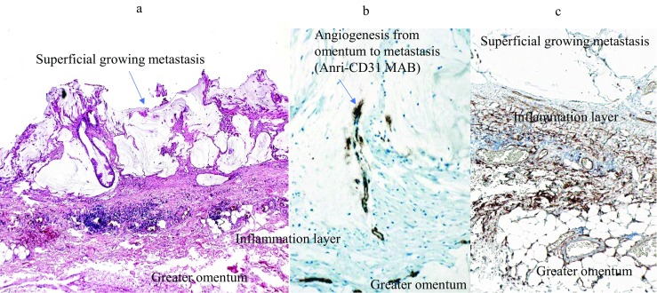
Fig. 5.

Microscopic findings of superficial growing metastasis. Tumor grows on a layer of the inflammatory cells on the greater omentum (A). Stromal tissue in the inflammatory layer shows positive immunoreaction against anti-CD34 monoclonal antibody (Mab) (C). Newly formed immature blood vessels, stained with anti-CD31 Mab, extended into the superficial growing metastasis from the preexisting blood vessels in the greater omentum (B).
