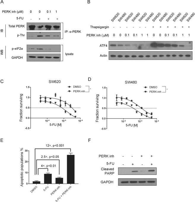
Figure 3.
Chemical inhibition of PERK sensitizes colon cancer cells to 5-Fluorouracil treatment. (A) Total PERK protein was immunoprecipitated from cells as indicated, and western blotting was conducted to measure the level of phosphorylated PERK by use of a pan-pThr/Ser antibody. Cellular level of p-eIF2α was also measured for cells treated as indicated. (B) Western blotting showing the effects of an escalating dose of PERK inhibitor (0 µM, 0.1 µM and 1 µM) on ATF4 in SW620 and SW480 cells w/or w/o Thapsigargin treatment. β-Actin was used as loading control. (C,D) Dose response curves showing effects of the PERK inhibitor (1 µM) in sensitizing SW620 and SW480 cells to 5-FU treatment. (E) Flow cytometric analysis showing the percentage of apoptotic cells (SW620) treated as indicated. (F) Western blot showing the protein level of cleaved PARP which reflects the degree of cell apoptosis (SW620) treated as indicated. * means P < 0.05. Data are represented as mean ± SEM or the mean alone.

