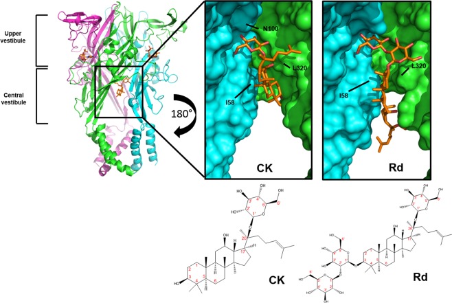
Figure 1.
Ginsenosides bind to the central vestibular region of P2X7. Representation of a homology model of hP2X7 trimer in the open state with ginsenoside CK docked in the central vestibular region. Each subunit is differentially coloured (green, cyan, magenta). Panels show a zoom-in and rotated view of the binding site from the inside of the cavity facing outwards (rotation of 180°). Surface rendered images are shown to visualise the binding pocket for CK and Rd on hP2X7. Ligands and side chains are represented as sticks with all hydrogen atoms omitted for clarity. Two-dimensional chemical structures of CK and Rd are also displayed with carbon atoms labelled in red.

