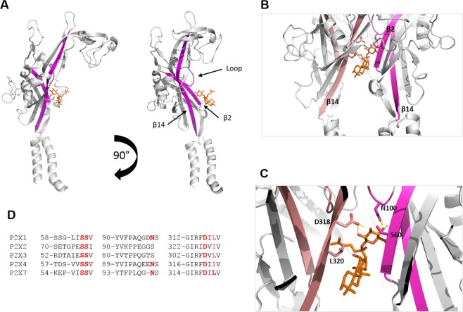
Figure 2.
Ginsenoside binding pocket consists of interactions with D318, N100 and S60 residues in hP2X7 lower body region. (A) An individual subunit is displayed with two lower body region β-sheets highlighted in magenta (β2 and β14). CK is shown docked into the putative binding site. Rotation of this individual subunit by 90° allows better visualisation of the internal loop containing N100 that is predicted to make contact with the glucopyranose ring of ginsenoside CK. (B) Trimeric view of the hP2X7 model showing the docked ginsenoside connecting inter-subunit β-sheets β2 (magenta) and β14 (salmon pink). (C) Closer view of the docked ginsenoside CK displaying interacting residues predicted to make polar contacts; D318, N100 and S60.

