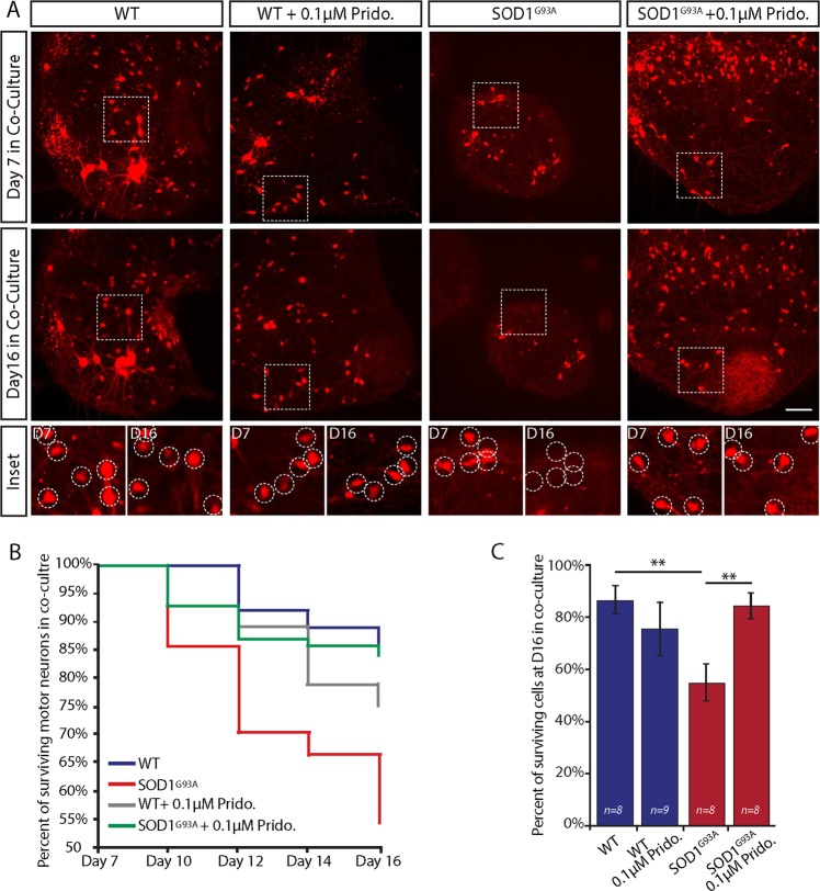
Fig. 2. Pridopidine impedes motor neuron loss in co-cultures of SOD1G93A.
a Representative images of either pridopidine treated or control SOD1G93A/ChAT::tdTomato and WTChAT::tdTomato spinal cord explants in the proximal compartment of MFCs in co-culture. Upper panel shows the ChAT-positive motor neurons (red) within each spinal cord explant at day 7 of co-culture. Central panel shows images of the same spinal cords at day 16 of co-culture. Bottom panel includes magnified images of the regions from the upper panels denoted by a dashed line. Images display the massive death of motor neurons in the SOD1G93A explants in co-cultures, whereas the number of surviving motor neurons in the WT and pridopidine-treated SOD1G93A remains roughly the same. Dashed circles denote motor neurons. b Plot of the average percentage of surviving motor neurons per explant over time displays accelerated motor neuron death in the SOD1G93A co-cultures that is profoundly impeded by 0.1 µM pridopidine application. c Bar chart showing the percentage of surviving cells per explant at day 16 in co-culture, which reveals a substantial rescue effect for pridopidine on SOD1G93A motor neurons. Data are shown as mean ± SEM. **p value < 0.01. (n = number of spinal cord explants from 3 or more independent experiments; Student’s t test.)

