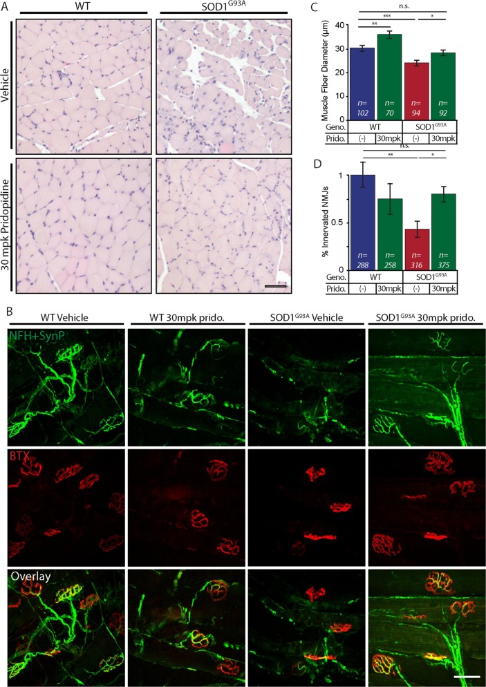
Fig. 7. Pridopidine ameliorates muscle fiber wasting and preserves NMJs in 16-week-old SOD1G93A mice.
a Representative images of H&E-stained cross-sections from Gastrocnemius muscle of mice from 4 groups: WT-vehicle, WT-30mpk pridopidine, SOD1G93A-vehicle, and SOD1G93A-30mpk pridopidine. The muscle histology of SOD1G93A-vehicle mice is poor and reveals a smaller diameter of muscle fiber, compared with WT-vehicle and WT-30mpk pridopidine. Muscles from pridopidine-treated SOD1G93A mice exhibit a larger and more homogenous muscle fiber population that resembles WT muscles. b Representative confocal images of muscle whole-mount preparations from 16-week-old mice stained for Postsynaptic AchR (BTX; red) and Presynaptic NFH + Synapsin-I + Synaptophysin (green). Muscles of SOD1G93A vehicle-treated mice show the degeneration of presynaptic neurons and the amorphous post-synaptic apparatus. SOD1G93A-30mpk pridopidine-treated mice display co-localized pre- and post-synaptic markers with a typical healthy NMJ morphology. c Quantitative analysis of muscle fiber diameter indicates that SOD1G93A-vehicle-treated muscle fibers are significantly smaller in diameter than are WT-vehicle fibers. 30mpk of pridopidine led to a significant ~4 µm increase in the muscle fiber diameter in SOD1G93A and ~5 µm in WT muscles. (n = number of muscle fibers.) d Quantitative analysis of the percentage of innervated NMJs reveals a massive ~60% loss of NMJs in the SOD1G93A-vehicle group, compared with the WT-vehicle group. Pridopidine treatment limited the loss of NMJs in SOD1G93A mice to ~20%. Data are shown as mean ± SEM (n = number of NMJs). *p value < 0.05; **p value < 0.01; ***p value < 0.001 (n = 5 mice in each group; Student’s t test)

