Figure 2.
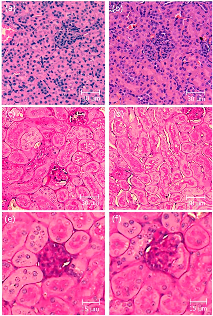
Histopathological analysis of kidney sections. Representative pictures of kidney sections stained by hematoxylin/eosin (H&E) (a,b) and periodic acid-Schiff (PAS) (c–f). Kidneys from control receiving vehicle (a, c and e) and crotamine-treated (b,d and f) mice. The glomerulus structure is shown in the magnified images (e and f). No evidence of histopathological lesions was noticed in the kidney sections analyzed by trained pathologists. Bar = 30 μm (a–d) and 15 μm (e,f).

