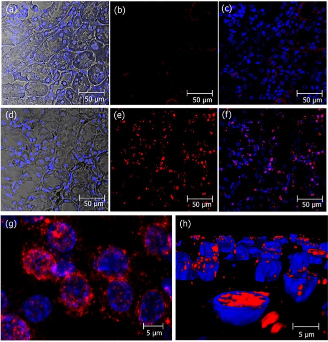
Figure 3.
Localization of crotamine in kidneys PTECs of mice receiving daily crotamine by ip injections during 21 days. Kidney sections from mice receiving vehicle saline (a–c) or native crotamine (1 µg/animal), daily, for 21 days (d–h). The perinuclear localization of crotamine (red) is shown in the magnified picture (g). The presence of crotamine (red) inside the nuclei was confirmed by the three-dimensional reconstitution of cell nucleus as shown in (h). Differential interference contrast (DIC) with nuclei stained with DAPI (blue) (a and d), and crotamine immunorecognized by rabbit anti-crotamine antibody followed by signal amplification by the Tyramide Signal Amplification (TRITC-labeled tyramide solution, tetramethyl rhodamine system, in red) (b,c,e,f,g and h). Overlay of red and blue fluorescence (c,f,g and h). Bar = 50 μm (a–f) and 5 μm (g, h).

