Figure 4.
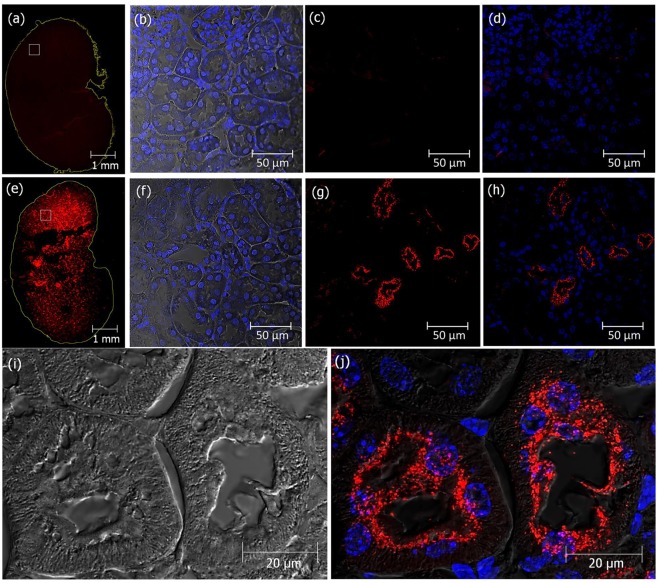
Cy3-labeled crotamine is reabsorbed from pro-urine by PTECs. Kidney sections from mice receiving vehicle (a–d) or a single ip administration of fluorescently-labeled Cy3-crotamine (5 µg/animal), 2 h before animal euthanasia (e–j). Fluorescence microscopy of transversal section of whole kidney (a and e), in which the boxes correspond to the areas magnified (as presented in b,c,d,f,g and h). Zoom out of kidney transversal section of mice showing the presence of Cy3-crotamine in the brush borders area of PTECs (i,j). Differential interference contrast (DIC) showing kidney tissue structure with nuclei stained with DAPI (blue) (b,f), and Cy3-crotamine stained in red (a,c,d,e,g,h and j). Differential interference contrast (DIC) image (i), and overlay of red and blue fluorescence with DIC (d,h and j). Bar = 1 mm (a,e), 50 μm (b–d, f–h), and 20 μm (i, j).

