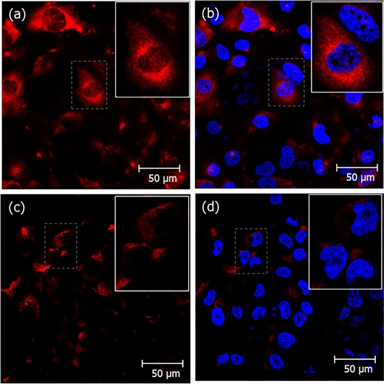
Figure 6.
Reduced crotamine uptake in Synd-1 deficient HK-2 cells. Wild-type human PTEC HK-2 cells (a,b) and Synd-1 KD cells (c,d) treated with crotamine (5 μM) for 1 h, at 37 °C. Nuclei were stained with DAPI (in blue), and crotamine recognized by antibody after signal amplification with TSA-TRITC (in red). Crotamine (red) is visualized in the cytoplasm of wild-type human PTEC HK-2 cells, whilst in Synd-1 KD cells, significantly reduced fluorescence is noticed. Bar = 50 μm.

