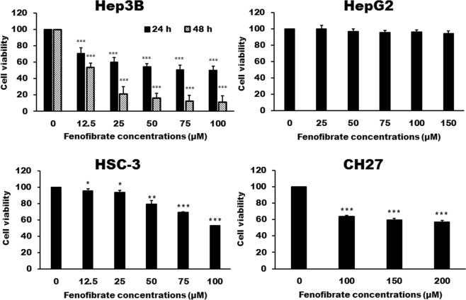
Figure 2.
Cytotoxicity of fenofibrate in Hep3B, HepG2, HSC-3 and CH27 cells. Hep3B, HepG2, HSC-3 and CH27 cells were incubated with vehicle alone or with various concentrations of fenofibrate for 24 h. The cytotoxicity was assessed by Trypan blue exclusion assay and the viable cells were counted. The data are expressed as the mean percentage of control ± S.D. of four independent experiments performed in triplicate. *P < 0.05, **P < 0.01, ***P < 0.001 compared to the control values.

