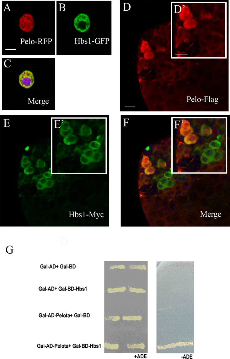
Figure 5.
Pelo physically interacts with Hbs1. (A–C) Co-expression of Pelo (A, Pelo-RFP, red) and Hbs1 (B, Hbs1-GFP, green) in Drosophila S2 cells, where those two proteins were co-localized in the cytoplasm (C: merged image with DAPI in blue). (D–F) UASp-Pelo-flag (D, red) and UASp-Hbs1-myc (E, green) were both expressed in the cytoplasm of testicular germ cells when they were driven by nosGal4. Note that they could co-localize with each other in the germline cells (F). (G) Pelo interacted with Hbs1 in a yeast two hybrid assay. Scale bar represents 10 μm in A–C and 15 μm in D–F.

