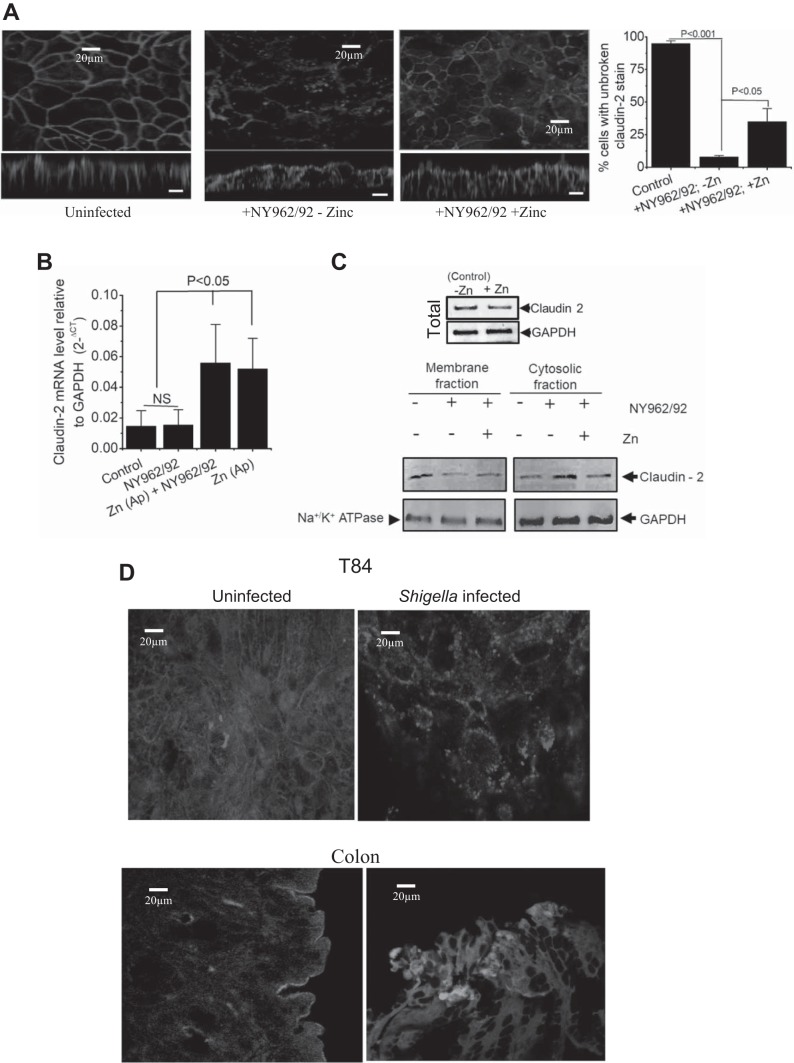
Fig. 4.
Altered distribution of claudin-2 is rescued by Zn2+ for restoration of paracellular permeability and barrier tightens. A: representative images of claudin-2 (red) and apical membrane marker wheat germ agglutinin (blue) in control (uninfected) and Shigella infected in the presence (NY962/92 + zinc) and absence (NY962/92 – zinc) of Zn2+ in T84 monolayer. Claudin-2 staining at cell-cell contacts is shown diminished by Shigella infection (XY). X-Z images showed that claudin-2-positive red strip in the apical region between neighboring cells were disturbed (NY962/92 – zinc) but relocalization of claudin-2 occurs in the presence of Zn2+ (NY962/92 + zinc). Bar graph represents quantification of multiple images based on continuity of claudin-2 stain (broken vs. unbroken). Three fields of view from 2 independent experiments for each condition, ~75 cells were scored. B: qRT-PCR showed that claudin-2 mRNA is markedly elevated by Zn2+ application in T84 cells. Data are means ± SE; n = 3 per group. C: effect of Shigella infection in presence or absence of Zn2+ on claudin-2 level in cytosolic and membrane fractions of T84 cells. Representative Western blot presented represent 3 independent experiments. Na+-K+-ATPase and GAPDH were used as loading control. Top: representative Western blot for claudin-2 abundance in presence and absence of Zn2+ without Shigella infection. D: T84 monolayers (top) and mouse distal colon (bottom); uninfected (left) or Shigella infected (right) were immunolabeled with anti-cystic fibrosis transmembrane conductance regulator (CFTR) antibody. Each image is representative of 3 separate experiments performed in duplicate.

