Fig. 2.

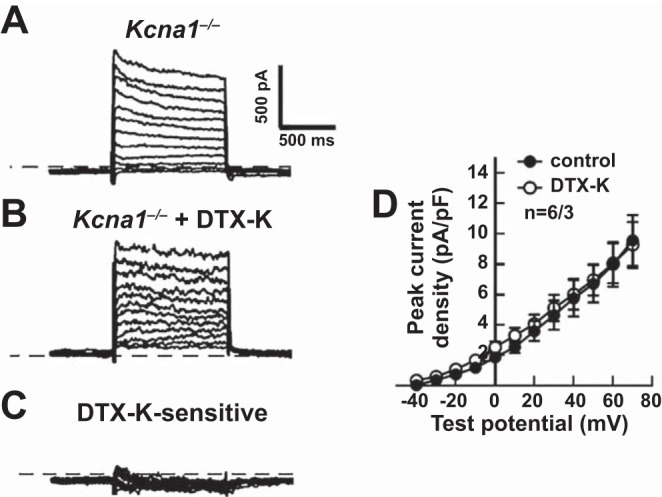
Dendrotoxin-K (DTX-K) had no significant effect on outward K+ currents in Kcna1−/− atrial myocytes. A–C: representative raw current traces in a Kcna1−/− cell in response to 1-s depolarizing voltage steps of +10 mV from a holding potential of −50 mV to +70 mV: at baseline (A); with application of 10 nM DTX-K (B); and following subtraction of the current difference to isolate the DTX-K-sensitive component (C). D: quantification of the peak current densities before (control) and after (DTX-K) application of DTX-K in Kcna1−/− atrial myocytes. The dotted line in the traces indicates 0 pA. Sample numbers (n) indicate myocytes per mouse.
