Fig. 1.
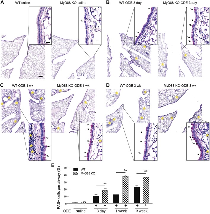
Myeloid differentiation factor 88 (MyD88)-deficient mice have reduced lung inflammation and increased airway periodic acid Schiff (PAS) staining following organic dust extract (ODE)-mediated inflammation. Mice were challenged with either intranasal saline for 3 days or intranasal 12.5% ODE for 3 days, 1 wk (8 airway challenges), or 3 wk (15 airway challenges; A–D). The day of the last challenge, mouse lungs were fixed with formalin and stained by PAS. Insets: low-power images were shown with higher magnification images. Arrows indicate PAS-positive cells. Yellow asterisks indicate regions of inflammatory cell infiltrate. Original scale bar, 100 μm for low-power images and 20 μm for high-magnification insets. E: quantification of percent PAS-positive cells per airway for each group (n = 1 for saline mice, and n = 2 mice for ODE-challenged groups, with 10–13 airways per mouse). **P < 0.05, significant difference between wild-type (WT) and MyD88 knockout (KO) mice.

