Fig. 5.
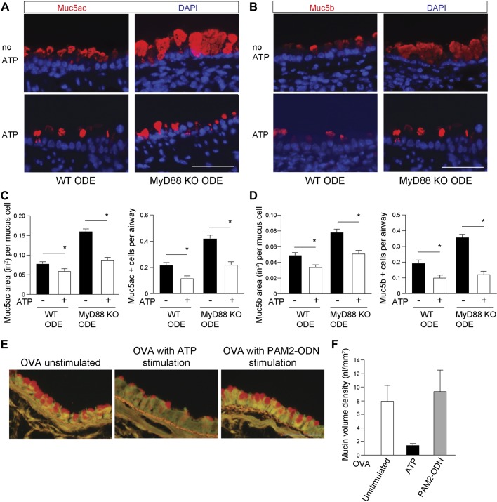
Myeloid differentiation factor 88 (MyD88) is not required for ATP-activated mucin secretion in vivo. Representative images of mucin (Muc)5ac detected by fluorescent-labeled Ulex europaeus agglutinin I lectin (A) and Muc5b with mouse MAb (B) in organic dust extract (ODE)-challenged wild-type (WT) and MyD88 knockout (KO) mouse airways. Bottom: representative images of mucin staining, followed by ATP in vivo stimulation. Top: images of staining without ATP (no ATP). Bar graphs depict mean value and SE of Muc5ac (C)- and Muc5b (D)-staining area and number of positive mucous cells per airway, with and without ATP stimulation. Values for no ATP are taken from Fig. 2 quantification (n = 5 mice per condition; 8–12 images per mouse). Original scale bars, 50 μm. *P < 0.05, significant difference between ODE and saline challenges. E: mucous metaplasia was induced by intraperitoneal immunization to ovalbumin (OVA), followed by aerosol exposure to OVA. Mice were then exposed to aerosolized ATP or Pam2CSK (Pam2)-oligodeoxynucleotide M362 (ODN) to induce mucin secretion, then mice were euthanized after 30–60 min, and excised lungs were stained with fluorescent periodic acid Schiff to examine intracellular mucin content. Representative images are shown of mice with OVA-induced mucous metaplasia but not challenged with an aerosolized secretagogue (left), challenged with ATP (middle), or challenged with Pam2-ODN (right). F: bar graphs depict mean values and SE. DAPI, 4′,6-diamidino-2-phenylindole.

