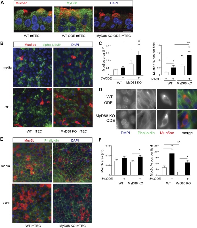
Fig. 8.
Organic dust extract (ODE) increase in mucin (Muc)5ac is augmented on myeloid differentiation factor 88 (MyD88)-deficient airway epithelial cells in vitro. A: MyD88 immunostaining (green) with Ulex europaeus agglutinin I (UEA-1) localization of Muc5ac (red) of mouse tracheobronchial epithelial cell (mTEC) isolated from wild-type (WT) and MyD88 knockout (KO) mice and grown under air–liquid interface (ALI) conditions ±5% ODE for 5 days. Original scale bar, 10 μ. B: representative images by UEA-1 staining for Muc5ac and acetylated α-tubulin for cilia in WT or MyD88-KO mTECs under ALI conditions challenged ±5% ODE for 5 days. Original scale bar, 50 μm. C: quantification of Muc5ac-staining area and percent-positive Muc5ac per cellular field from B (n = 4 per condition with 5 images per sample). D: representative higher magnification images demonstrating intracellular UEA-1- Muc5ac (red) staining with phalloidin (green) to mark plasma membrane borders. Original scale bar, 10 μm. E: representative images of Muc5b immunostaining with phalloidin to mark plasma membrane borders in WT or MyD88-KO mouse-derived mTEC under ALI conditions as, in part, A. F: quantification of Muc5b staining from A (n = 3 per condition with 5 images per sample condition). *P < 0.05, significant difference between ODE and saline. **P < 0.05, significant difference between WT and MyD88-KO mice. DAPI, 4′,6-diamidino-2-phenylindole.

