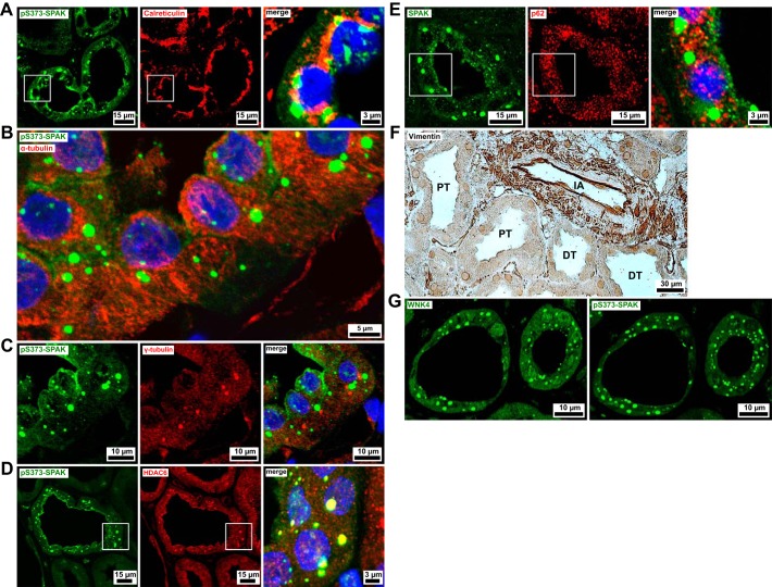
Fig. 2.
Inclusions of Na+-Cl− cotransporter (NCC)-activating kinases are found adjacent to the endoplasmic reticulum and microtubules. All images in this figure are from biopsies of patients with chronic hypokalemia. A: the endoplasmic reticulum lumen protein calreticulin is consistently localized in the vicinity of puncta labeled by antibody against residues 367–379 of human Ste20-related proline-alanine-rich kinase (SPAK) phosphorylated on Ser373 (pS373-SPAK) puncta in the perinuclear compartment. B: pS373-SPAK-positive puncta are arranged alongside filamentous α-tubulin (microtubular) immunofluorescence. C: pS373-SPAK does not accumulate around microtubule organizing centers (MTOCs). MTOCs are visible as sites of concentrated γ-tubulin expression, appearing once per cell. D: Histone deacetylase 6 (HDAC6) is expressed in all renal segments and compartments. In the distal convoluted tubule (DCT), it displays an intense, punctated signal colocalized with pS373-SPAK. E: p62 shows a speckled cytoplasmic pattern in all renal segments and compartments, with no apparent spatial relationship to the pS373-SPAK signal in the DCT. F: immunoperoxidase staining of vimentin is limited to interstitial and vascular compartments, whereas tubular epithelia are vimentin-negative (DT, distal tubule; IA, interlobular artery; PT, proximal tubule). G: the hypokalemic DCT contains punctate with no lysine kinase-4 (WNK4) accumulations. WNK4 immunostaining was performed after eluting several other primary antibodies. In the first round of staining, pS373-SPAK was detected in equally located cytoplasmic puncta. Blue channels show DAPI nuclear counterstain.

