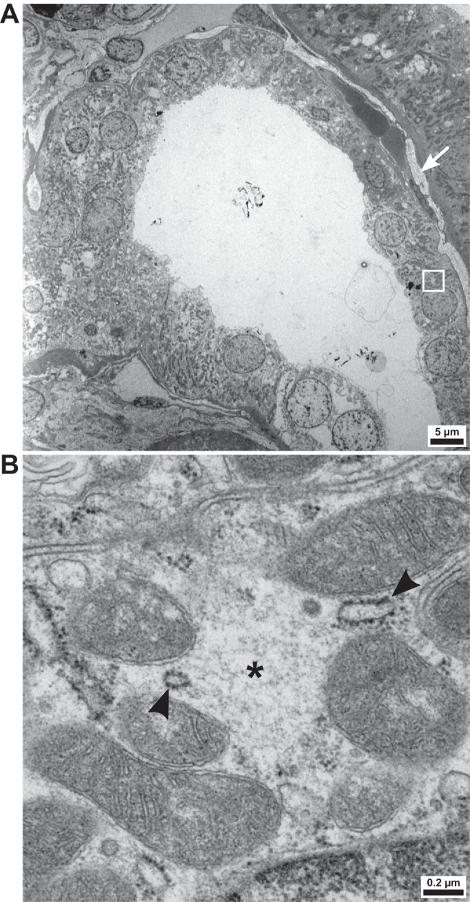
Fig. 3.

Ultrastructure of the distal convoluted tubule (DCT) of a patient with hypokalemia. A: overview of a cross section of the DCT in the patient with Munchausen syndrome. The region of interest (see B) is indicated by a white rectangle. To the right of the tubule, interstitial collagen deposits are visible (arrow). B: the DCT is enriched with hypodense, spherical cytoplasmic regions not delineated by membranes (*). Their ultrastructure is compatible with prior descriptions of with no lysine kinase (WNK) bodies. Rough endoplasmic reticulum (arrowheads) is found at their borders.
