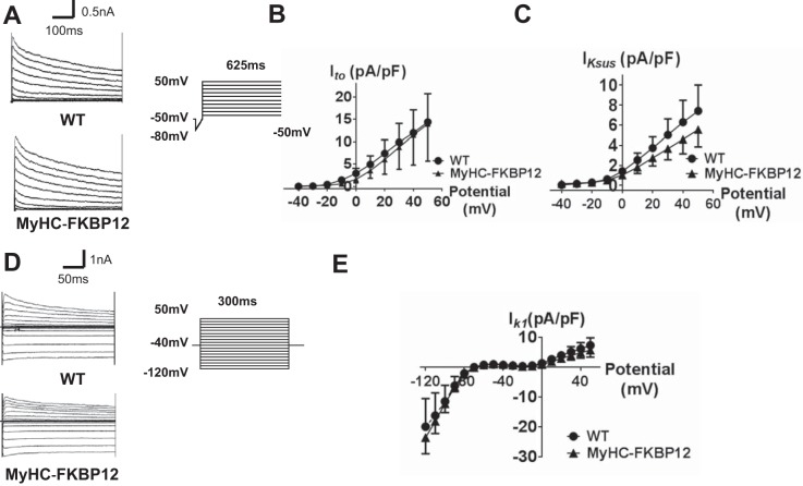
Fig. 6.
Analysis of cardiac transient outward K+ currents (Ito), sustained K+ currents (IKsus), and inward rectifier K+ currents (IK1) from isolated atrial cardiomyocytes in nontransgenic (NTG) and FK506-binding protein 12 transgenic (αMyHC-FKBP12) mice. A: representative Ito traces of NTG and αMyHC-FKBP12 myocytes. Ito was elicited from a holding potential at −50 mV with a 50-ms ramp from −80 to −40 mV followed by depolarizing pulses from −40 to +50 mV for 625 ms (in 10-mV increments). B and C: current-voltage (I-V) relationship (I-V curve) for Ito and IKsus. NTG: n = 21 cells/4 mice and αMyHC-FKBP12: n = 14 cells/4 mice. D: representative IK1 traces of NTG and αMyHC-FKBP12 myocytes. IK1 was elicited by holding the cell at −40 mV followed by 10-mV steps from −120 to 50 mV for 300 ms. E: I-V curve for IK1. NTG: n = 22 cells/4 mice and αMyHC-FKBP12: n = 18 cells/4 mice. WT, wild type.

