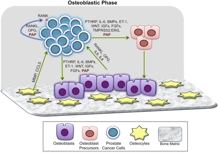
Figure 2.
Key players in the osteoblastic phase of prostate cancer bone metastases. In the osteoblastic phase, PCa cells continue to interact with all of the components of bone, although paracrine (green arrow) and autocrine (blue arrow) mechanisms as noted. A number of PCa-derived soluble factors such as ET-1, WNTs, TGF-β, uPA, IGF-1, FGFs, and BMPs have been shown to induce the dysfunctional osteoblastic phenotype. High levels of OPG are associated with end-stage OB bone metastases. PAP secreted by PCa cells in bone has both autocrine and paracrine effects that coordinately result in higher OPG/RANKL in the bone niche, resulting in osteoblastic lesions. ET-1, endothelin-1.

