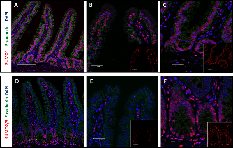
Figure 1: SUMO1 and SUMO2/3 distribution in murine small intestine.
Immunostaining of small intestinal sections from animals that were paraformaldehyde-perfused via transcardiac puncture. Sections were stained as indicated with e-cadherin (green), DAPI (blue), and either SUMO1 (red: A-C) or SUMO2/3 (red: D-F). A and D: small intestinal section, 200x; B and E: villus tip, 600x; C and F: crypt, 600x.

