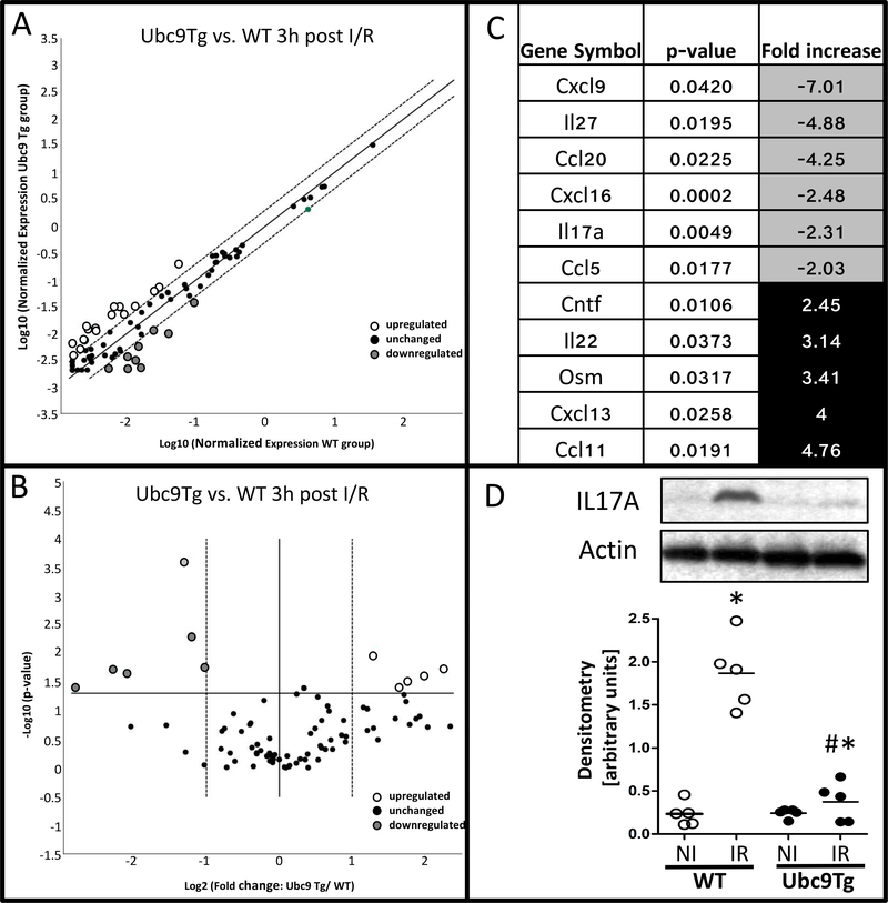
Figure 5: Modulation of villus-tip epithelial responses in Ubc9Tg animals after I/R.
Intestinal epithelial villus-tip epithelia from ischemic regions of wild type and Ubc9Tg (45 minutes of ischemia and 3 hours of reperfusion) were isolated by laser microdissection and capture and RNA was isolated to perform a cytokine targeted PCR array. A) Scatter and B) Volcano- blot outline the distribution of regulated genes, significantly up- and downregulated genes are listed in C). D) IL17A protein levels in non-ischemic small intestinal loops (NI) and loops subjected to ischemia/reperfusion (IR; 45minutes ischemia and 6 hours or reperfusion). Quantification was performed by densitometry and is represented as the ratio of IL17A to bActin band intensity. n=5; *p≤0.05

