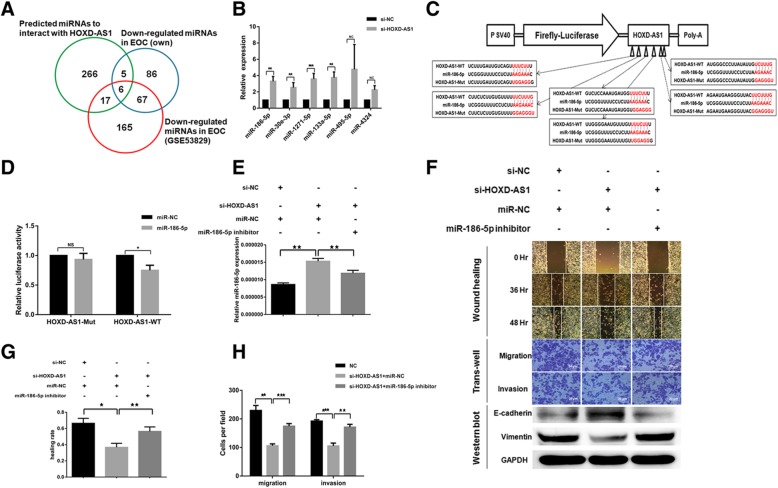
Fig. 4.
HOXD-AS1 binds to miR-186-5p to promote EOC migration, invasion and EMT. a Venn diagram showing the six overlapped miRNAs which were significantly down-regulated in the Affymetrix miRNA arrays and GSE53829, and predicted to be a target of HOXD-AS1. b Relative expression of the six miRNAs in (a) in EOC cells transfected with siRNA against HOXD-AS1 or si-NC control, measured using RT-qPCR. c Schema showing the cloning of wild type as well as a mutant HOXD-AS1 in which all six putative miR-186-5p binding sites were mutated in a luciferase reporter construct. d Direct interaction of HOXD-AS1 and miR-186-5p on the putative miR-186-5p binding sites as shown by the specific reduction of relative luciferase reporter activities in cells co-transfected with miR-186-5p and wild type HOXD-AS1 reporter construct. e miR-186-5p expression was elevated in EOC cells transfected siRNA against HOXD-AS1 and this could be reversed by the co-transfection of miR-186-5p inhibitors, as shown in RT-qPCR with U6 snoRNA normalization. ★ denotes p < 0.05, ★★denotes p < 0.01. f EOC cells transfected with si-HOXD-AS1 showed reduced ability to migrate and invade in vitro as measured through wound healing and trans-well assays (imaged at × 200 magnification) than control cells, with a corresponding increase in E-cadherin (an epithelial marker) and decrease in Vimentin (a mesenchymal marker). These changes in phenotypes could be partially rescued with the co-transfection of miR-186-5p inhibitors which partially reversed the miR-186-5p elevation by HOXD-AS1 knock-down, suggesting HOXD-AS1 mediated these phenotypic changes through miR-186-5p (quantitation graph of WB refer to Additional file 3: Figure S2C). g, h Relative wound healing rate (g) and transwell migration/invasion(h) from at least three independent experiments as described in(f)

