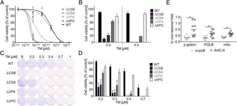
Fig. 4.
TM induces hypersensitivity in human DNA repair-deficient cells and causes DNA lesions in vivo. (A) Dose–response survival curves of TM-treated mutants deficient in CSA, CSB, and XPA, but not XPC, show TM hypersensitivity compared with wild type (WT). Values are normalized to solvent controls and represent means ± SEM of three technical replicates. Data from one of three biological replicates are shown. (B) Cell viability shown as means ± SEM at two assay concentrations (indicated with arrows in A). (C) Colony formation by cells treated with n-butanol (B) or the indicated concentrations of TM before recovery in drug-free medium. Macroscopic colonies were stained with crystal violet. (D) Values of C normalized to solvent. Means ± SEM are shown (n = 3). Significance of results for mutants compared with WT was determined with one-way ANOVA followed by Sidak’s multiple comparison (*P ≤ 0.05). (E) Higher lesion burden in genomic DNA isolated from cecal enterocytes of K. oxytoca AHC-6 (WT) colonized mice (24 h) compared with mice infected with the ∆npsB-mutant was detected with long amplicon quantitative PCR. Lesions/10 kb in β-globin, DNA-polymerase β (POLB) and in a region of the mitochondrial genome (mito.) are shown. Bars indicate means (n = 9), and significance was determined with unpaired t test (*P ≤ 0.05).

