Fig. 6.
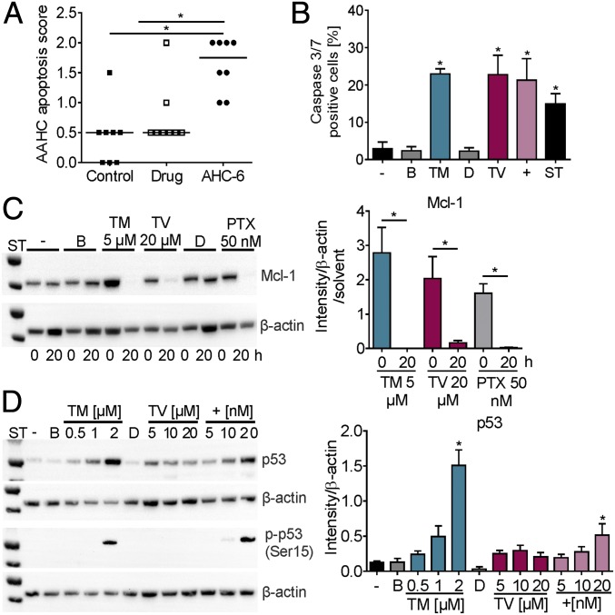
In vitro effects of TM and TV match features of AAHC. (A) Histopathological apoptosis scores of colons of untreated (control), amoxicillin/clavulanate treated (drug), and mice colonized with K. oxytoca (AHC-6). Bars indicate medians (n = 8). Kruskal–Wallis test followed by Dunn’s multiple comparison (*P ≤ 0.05). (B) Percent caspase 3/7-positive SW48 cells without (−) or after 24-h treatment with 2 µM TM, 60 µM TV, 20 nM GWL-78 (+), 2 µM staurosporin (ST), and solvents (B and D). Values are means ± SD (n = 3). (C) Total Mcl-1 protein in HT-29 cells treated 0 and 20 h with TM, TV, PTX, or solvents. Means ± SEM of signal for total Mcl-1 normalized to β-actin and solvents obtained from three independent cell lysates are shown (Right). (D) Total p53 protein and Ser15 phosphorylation (p-p53) detected in SW48 lysates treated 12 h with TM, TV, and GWL-78 (+) as indicated (Left). Means ± SEM of signal for total p53 normalized to β-actin obtained from three independent cell lysates are shown (Right). One-way ANOVA followed by Sidak’s multiple comparison (*P ≤ 0.05).

