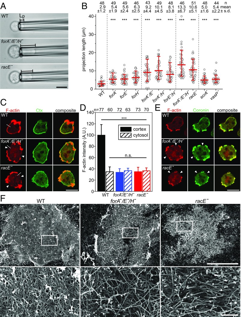
Fig. 4.
Elimination of the three cortical formins or of RacE increasingly impairs cortical rigidity and disrupts the contractile actin cortex. (A) Lp of WT and the mutant cells indicated was determined by micropipette aspiration using a constant suction pressure of 500 Pa from time-lapse movies; data correspond to Movie S2. (Scale bar, 10 µm.) (B) Quantitative analysis of the projection lengths of probed cells. ***P < 0.001 by Mann–Whitney rank sum test. n, number of analyzed cells. Statistical differences refer to WT. (C) Defects of the actin cortex in forA−/E−/H− and racE− mutants. Fixed WT and mutant cells were stained with a monoclonal Ctx antibody (green) to visualize PI(4,5)P2-containing membranes and F-actin with phalloidin (red). The white arrow heads indicate regions of low cortical density in the mutants. (Scale bar, 10 µm.) (D) Quantification of cortical and intracellular actin in WT and mutant cells. Average intensity profiles along five-pixel-wide lines as shown in C by the white dashed lines. n, number of analyzed cells; error bars, SDs. (E) Prominent F-actin assemblies outside the cortical breaches in forA−/E−/H− and racE− cells are identified as protrusions containing coronin (green), particularly marking Arp2/3 complex-driven actin networks, and phalloidin-stained F-actin (red). (Scale bar, 10 µm.) (F) Representative SEM micrographs of detergent-extracted WT and mutant cells (low magnification overviews on Top and high magnification of Insets at Bottom, respectively). [Scale bars: overview, 5 µm; Insets, 0.5 µm.]

